3D Reconstruction of Zebrafish larvea using VAST
These pages are in support of:
3D Reconstruction and Measurement of Zebrafish Larvea from High-Troughput axial-view in vivo imaging.
Yuanhao Guo, Wouter J. Veneman, Herman P Spaink, Fons J. Verbeek (2017)
BioMedical Optics Express 8(5): 2611-2634
We have modelled 3 stages of zebrafish larvae form axial views generated with our VAST-BioImager.
- Model 1, 3 dpf Zebrafish larvae
- Model 2, 4 dpf Zebrafish larvae
- Model 3, 5 dpf Zebrafish larvae
In order to obtain the models, we have used a Hybrid Segmentation method that we have developed to overcome segmentation errors that result from the application of standard methods. A summary of the method can be seen here [LINK].
The animations represents voxel models as they are reconstructed with our methods from profiles obtained from segmentations of axial-views.
The following animations illustrate three different models produces with our reconstruction methods.
The viewer can appriciate the correctness of the reconstruction for each of the larval stages.
Animation 1
Here we see the reconstruction of a 3df zebrafish larva from VAST.
| Input Axial Image (n=21 views) & Segmentation | |
| 3 dpf | 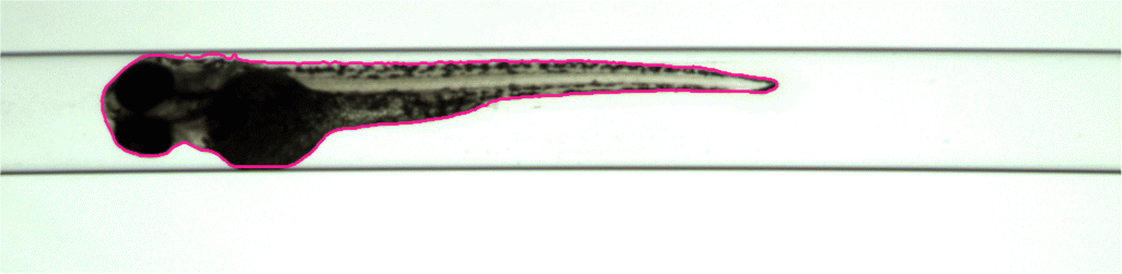 |
| 3D model | |
| 3 dpf | 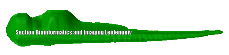 |
| 3D model with texture mapping | 3 dpf | 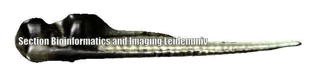 |
Animation 2
Here we see the reconstruction of a 4dpf zebrafish larva from VAST.
| Input Axial Image (n=21 views) & Segmentation | |
| 4 dpf | 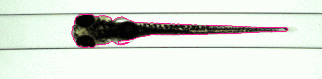 |
| 3D model | |
| 4 dpf | 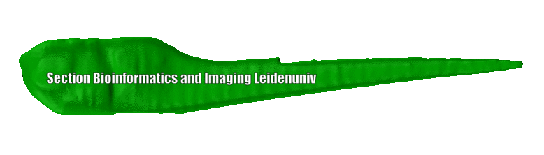 |
| 3D model with texture mapping | 4 dpf | 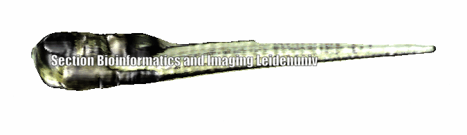 |
Animation 3
Here we see the reconstruction of 5 dpf zebrafish larva from VAST.
| Input Axial Image (n=21 views) & Segmentation | |
| 5 dpf | 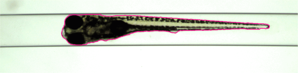 |
| 3D model | |
| 5 dpf | 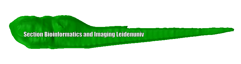 |
| 3D model with texture mapping | 5 dpf | |
Contact Yuanhao Guo or Fons Verbeek for information.