Gallery pages for the European Pond Turtle (Emys orbicularis)
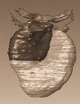
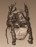
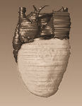
Modelling anatomy and development of the Emys orbicularis heart, other stages
This page gives access to a collection of additional pictures (under Pictures in the menu) of a number of canonical stages of heart development in Emys orbicularis. The Index in the menu links to the other developmental stages.
Legends: A, atrium; AA, aortic arch; ACV, anterior cardinal vein; AVC, atrioventricular canal; CA, cavum arteriosum; CAVV, cushion tissue forming the atrioventricular valve; CP, cavum pulmonale; CCV, common cardinal vein; CV, cavum ventrale; DC, distal cushions; F, foramen; HS, horizontal septum; HSt, heart stalk; IAS, interatrial septum; IHC, inner heart curve; IVC, intraventricular canal; LAA, left aortic arch; LA, left atrium; LAVC, left atrioventricular canal; MC, mesenchymal cap; OFT, outflow tract; PA, pulmonary artery; PC, proximal cushions; PCV, posterior cardinal vein; PM, pectinate muscle; PV, pulmonary vein; RAA, right aortic arch; RA, right atrium; SV, sinus venosus; TC, trabeculae carneae; V, ventricle; VS, vertical septum; VV, possible venous valve of the superior cardinal vein.
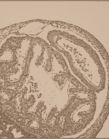
Histology.
In addition to the 3D reconstructions, histological sections were studied and described for Yntema's developmental stages 9 through 16. Here section images are shown in which the most interesting biological processes at different stages in cardiac development can be seen. For images on histology follow this link Histological sections.
Index to All Stages
 |
 |
 |
 |