Gallery pages for the European Pond Turtle (Emys orbicularis)
Anatomy and development of the Emys orbicularis heart, Yntema stage 15
This page gives access to a collection of additional pictures (under Pictures in the menu), animation sequences (under Animations in the menu) and interactive browsing using the 3D model browser (under Interactive in the menu) of heart development at stage 15 of Emys orbicularis. The Index in the menu links to the other developmental stages.
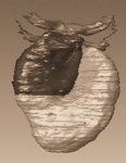
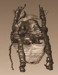
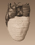
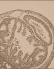
Legends: A, atrium; AA, aortic arch; ACV, anterior cardinal vein; AVC, atrioventricular canal; CA, cavum arteriosum; CAVV, cushion tissue forming the atrioventricular valve; CP, cavum pulmonale; CCV, common cardinal vein; CV, cavum ventrale; DC, distal cushions; F, foramen; HS, horizontal septum; HSt, heart stalk; IAS, interatrial septum; IHC, inner heart curve; IVC, intraventricular canal; LAA, left aortic arch; LA, left atrium; LAVC, left atrioventricular canal; MC, mesenchymal cap; OFT, outflow tract; PA, pulmonary artery; PC, proximal cushions; PCV, posterior cardinal vein; PM, pectinate muscle; PV, pulmonary vein; RAA, right aortic arch; RA, right atrium; SV, sinus venosus; TC, trabeculae carneae; V, ventricle; VS, vertical septum; VV, possible venous valve of the superior cardinal vein.
Yntema stage 15.
By now the distal bend of the original heart tube has reached the apex of the ventricle and the walls of both have merged. The caudal apex of the bend has formed the cavum pulmonale, continuous on the cranial end with the outflow tract. The merged wall between the cavum pulmonale and the rest of the ventricle has formed an incomplete septum, the horizontal septum. This is clearly visible in the cutaway views of the 3D model of this stage. The cavum pulmonale can be seen on the ventral side of the incomplete septum, at the base of the outflow tract, as a distinct cup-shaped chamber. It is almost completely separated from the rest of the ventricle, save for a small passage on the cranial side, connecting it to the cavum venosum. From this developmental series it can be concluded that the cavum pulmonale develops at the proximal end of the outflow tract and the horizontal septum is formed through bending of the tube. It is interesting to note that the cavum pulmonale has almost no trabeculae at this stage, while the rest of the ventricle is heavily trabeculated, with trabeculae five to seven times as high as they are wide. One of these trabeculae is slightly bigger with a dorso-ventral base thereby forming a septum at the apex of the ventricle. This is the vertical septum, dividing the cavum venosum from the much smaller cavum arteriosum. This septum is not as large as the horizontal septum. The atria and ventricle are no longer in open connection, but are separated by an atrioventricular canal, in which the atrioventricular cushions are suspended. The ventral atrioventricular cushion is still connected at one end to the proximal cushion tissue in the outflow tract and at the other end to the mesenchymal cap (as can be seen in the animation sequence of the cushion tissue, provided below). In the outflow tract the four distal and the two proximal cushions are now clearly visible. For images, follow this link: Developmental stage 15
Animations Yntema stage 15.
the complete model |
the cushion tissue |
Interactive Visualization
For Interactive Visualization and Manipulation of the Yntema Stage 15 model please use our 3D visualizer.
The interactive vizualization application is a java applet and it reads the model as produced in TDR-3Dbase (Verbeek, 1999). from our server. At the server the model is stored in a database which contains the section images, the structure information and the graphical information. The java applet requires Java 3D to be installed on your system and available for the browser. In case you cannot see the the 3D representation tab please contact your system administrator.The applet is preferably used over a high badwdtih internet connection; the computer should have a good graphics card and sufficient memory.
Instructions for correct use of the 3D browser and software that needs to be installed, click: [PDF]
To start interactive visualization click [HERE]
Index to All Stages
 |
 |
 |
 |