Gallery pages for the European Pond Turtle (Emys orbicularis)
Anatomy and development of the Emys orbicularis heart
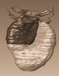
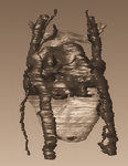
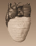
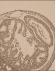
These pages provide additional images, animation sequences and interactive browsing of the 3D reconstructions, supplementing the Emys orbicularis project of LIACS, in collaboration with the Institute of Biology in Leiden (IBL), Leiden University. This research is presented in Bertens, L.M.F., Richardson, M.K., Verbeek, F.J. (2010)Analysis of cardiac development in the turtle Emys orbicularis (Testudines: Emidydae) using 3D computer modelling from histological sections; Anatomical Record 2010 (In Press). In this study the hearts of three embryos of this species were reconstructed in 3D computer models, in order to elucidate the embryonic development of the horizontal septum in the ventricle. Together with histological sections of the hearts these models comprise a developmental series. The models represent developmental stages 8, 10 and 15 as described by C.L. Yntema, (1968). Additional stages are described on the level of histology. For each of the 3D reconstructions interactive viewing, additional images and animation sequences are provided; of the histological study images provided.
 |
 |
 |
 |