Image acquisition for 3D reconstructions from serial sections
Serial sectioning techniques are frequently required in (developmental) biology to analyse and/or
describe phenomenona with a three-dimensional nature. However, when physically sectioning the biological
object of interest (e.g. an embryo), the three dimensional structure is lost and might be very hard to
envision. As an aid for analysis and visualization a 3D reconstruction can be made.
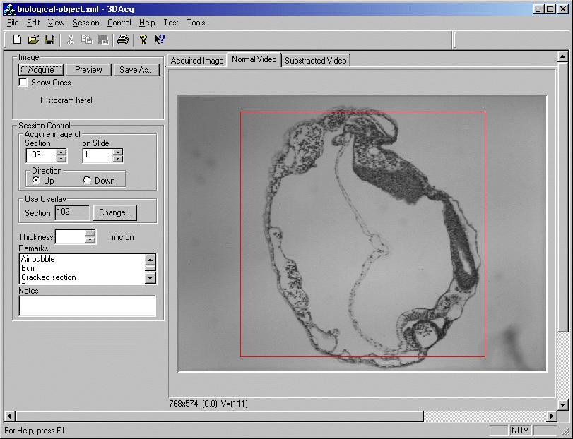
Using the 3D acquisition program we designed and implemented, users are able to acquire images of the
whole series of sections in an easy way. In this process not only image files are stored, but also other
information necessary for making a 3D reconstruction.
The following screenshot shows the latest version of our acquisition program.
Requirements for 3D acquisition
If you want to make a 3D reconstruction of your biological object some requirements need to be considered.
Preferably, your material should be embedded in plastic to have as less deformation from the sectioning
process as possible.
Another requirement is that your series is as complete as possible and that your sections are placed on
a glass slide in a specific order and position. For detailed information on our acquisition procedure: Fons Verbeek.
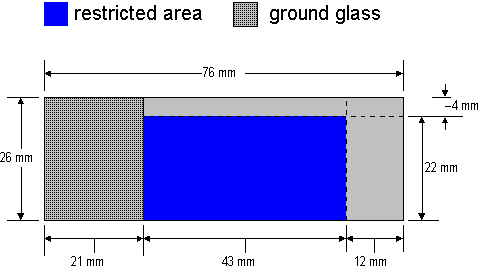
The sections should be placed on the restricted area of the object glass shown below.
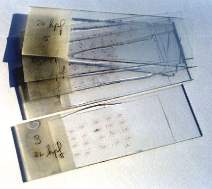
An example of a series of sections mounted on glass slides is given below.
Contact Fons Verbeek for information.